AG Hein: Mechanisms of neuronal and axonal degeneration in Multiple Sclerosis
Team Leader
Dr. Katharina Hein

- phone:
- + 49 - 551 - 39 14410
- pager:
- 919 - 1804
- e-mail:
- k.hein(at)uni-goettingen.de
Abstract
It is now widely recognized that axonal injury and consecutive neuronal degeneration play an important role in the pathogenesis of Multiple Sclerosis and that loss of axons is directly correlated with the irreversible progression of neurological deficits. Knowledge of the molecular mechanisms underlying neuro-axonal degeneration is therefore instrumental to develop suitable neuroprotective therapies for MS.
Major Research
Interest:
- Identification of the molecular mechanisms underlying inflammation-driven injury of the CNS
- Validation of the novel therapeutic targets
- Clinical focus: clinical studies in MS patients including investigator initiated clinical trials, MRI and MR spectroscopy, neuropsychological testing and CSF analysis
Tools and Models
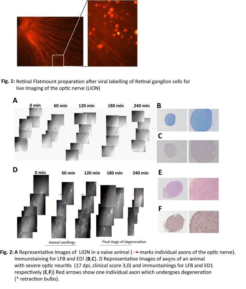
- In vivo: animal model of MS, optical coherence tomography, live imaging of axons, AAV-mediated gene transfer, acute and chronic infection models
- In vitro: T-cell culture, primary cell culture of retinal ganglion cells, molecular biology
 |
|
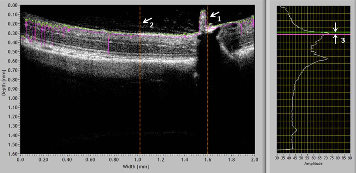
Figure: Optical coherence tomography in autoimmune optic neuritis: Measurements of RNFL thickness were performed in the region of interest (ROI) (500 µm temporal or nasal from the center of the optic disc (arrow 1)). RNFL thickness was determined as the distance (double arrow 3) between the two A-scan graphs (green and magenta lines) which correspond to the upper and lower boundary of the RNFL, respectively. |
 |
Selected original publications
- Sühs KW*, Hein K*, Sättler MB, Görlitz A, Ciupka C, Scholz K, Käsmann-Kellner B, Papanagiotou P, Schäffler N, Cordula Restemeyer C, Bittersohl D, Hassenstein A, Seitz B, Reith W, Fassbender K, Hilgers R, Heesen C, Bähr M, Diem R (2012). A randomized, double-blind, phase II study on erythropoietin in optic neuritis. Ann Neurol 72(2):199-210. *shared first authorship
- Hein K, Gadjanski I, Kretzschmar B, Lange K, Diem R, Sättler MB, Bähr M (2012) An optical coherence tomography study on degeneration of retinal fiber layer in rats with autoimmune optic neuritis. Invest Opthalmol Vis Sci 53(1):157-63.
- Gadjanski I, Williams SK, Hein K, Sättler MB, Bähr M, Diem R (2011). Correlation of optical coherence tomography with clinical and histopathological findings in experimental autoimmune uveitis. Exp Eye Res 93(1): 82-90.
- Rau CR*, Hein K*, Sättler MB, Kretzschmar B., Hillgruber C., McRae BL, Diem R. and Bähr M. (2011) Anti-inflammatory effects of FTY720 do not prevent neuronal cell loss in a rat model of optic neuritis. Am J Pathol 178(4):1770-81. *shared first authorship
- Hein K. (née Maier), Köhler A., Diem R., Sättler M.B., Demmer I., Lange P., Bähr M., Otto M. (2008): Biological markers for axonal degeneration in CSF and blood of patients with the first event indicative for multiple sclerosis. Neurosci Lett; 436(1):72-6.
- Maier K., Merkler D., Gerber J., Taheri N., Kuhnert A.V., Willciams S.K., Neusch C., Bähr M., Diem R. (2007) Multiple neuroprotective mechanisms of minocycline in autoimmune CNS inflammation. Neurobiol Dis; 25: 514-525.
- Maier K, Kuhnert AV, Taheri N, Sättler MB, Storch MK, Williams SK, Bähr M, and Diem R (2006) Effects of glatiramer acetate and interferon-beta on neurodegeneration in a model of multiple sclerosis: a comparative study. Am J Pathol 169: 1353-64.
- Diem R, Sättler MB, Merkler D, Demmer I, Maier K, Stadelmann C, Ehrenreich H, and Bähr M (2005) Combined therapy with methylprednisolone and erythropoietin in a model of multiple sclerosis. Brain 128: 375-385.
- Maier K, Rau CR, Storch MK, Sättler MB, Demmer I, Weissert R, Taheri N, Kuhnert AV, Bähr M, and Diem R (2004) Ciliary neurotrophic factor protects retinal ganglion cells from secondary cell death during acute autoimmune optic neuritis in rats. Brain Pathol 14: 378-387.
- Sättler MB, Merkler D, Maier K, Stadelmann C, Ehrenreich H, Bähr M, and Diem R (2004) Neuroprotective effects and intracellular signaling pathways of erythropoietin in a rat model of multiple sclerosis. Cell Death Differ 11: S181-192.
- Hobom M, Storch MK, Weissert R, Maier K, Radhakrishnan A, Kramer B, Bähr M, and Diem R (2004) Mechanisms and time course of neuronal degeneration in experimental autoimmune encephalomyelitis. Brain Pathol 14: 148-157.
- Diem R, Hobom M, Maier K, Weissert R, Storch MK, Meyer R, and Bähr M (2003) Methylprednisolone increases neuronal apoptosis during autoimmune CNS inflammation by inhibition of an endogenous neuroprotective pathway. J Neurosci 23:6993-7000.
The Team
Irina Graf, technician

- lab:
- + 49 - 551 - 39 12837
- office:
- + 49 - 551 - 39 20525
- e-mail:
- irina.graf(at)med.uni-goettingen.de
Techniques:
- Molecular biology
- Cell culture
- Histopathology
- Animal models
Prateek Kumar, PhD student

- lab:
- + 49 - 551 - 39 12837
- office:
- + 49 - 551 - 39 20525
- e-mail:
- singh_prats31(at)yahoo.co.in
Research Interest:
- Identification of genes and underlying molecular mechanism involved in early neurodegeneration in MOG-EAE
- Impact of chronic and acute infection on autoimmunity in MOG-EAE
Techniques:
- Chronic and acute infection models
- Microarray analysis
- Real time PCR
- Western Blot
- Histopathology
Johannes Ruhe, cand. med.

- lab:
- + 49 - 551 - 39 12837
- office:
- + 49 - 551 - 39 20525
- e-mail:
- johannes.ruhe(at)web.de
Research Interest:
Techniques:
- In vivo imaging
- Histopathology
- Animal models
Henrike Menkhoff, cand. med.

- lab:
- + 49 - 551 - 39 12837
- office:
- + 49 - 551 - 39 20525
- e-mail:
- henrikemenkhoff(at)yahoo.de
Research Interest:
- Spatio-temporal relation between immune attack and neuronal/axonal degeneration during EAE. Pathomechanisms of neurodegeneration.
Techniques:
- MOG-induced animal model of MS.
- Behavioral analysis of healthy and diseased animals (e.g. gait analysis)
- Histopathology